Monday, 15 April 2024

Understanding Slipped Capital Femoral Epiphysis (SCFE): Causes, Symptoms, and Treatment


Introduction:

Slipped Capital Femoral Epiphysis (SCFE) is a relatively rare but potentially serious hip disorder that mainly affects adolescents during their growth spurts. This condition occurs when the ball at the top of the thigh bone (femur) slips off the neck of the bone at the hip joint. Understanding SCFE is crucial for early detection and treatment to prevent long-term complications. In this article, we delve into the causes, symptoms, diagnosis, and treatment options for SCFE.


What Causes SCFE?

The exact cause of SCFE is not fully understood, but it is believed to be related to a combination of factors, including:

  1. Growth spurts: SCFE often occurs during periods of rapid growth, typically in pre-adolescents and adolescents. The growth plate at the top of the femur is weaker during these growth spurts, making it more susceptible to slipping.

  2. Obesity: Excess weight puts added pressure on the growth plate, increasing the risk of it slipping. Obese children are more likely to develop SCFE compared to those of normal weight.

  3. Hormonal changes: Hormonal changes during puberty may also contribute to the weakening of the growth plate, making it more prone to slipping.

  4. Genetics: There may be a genetic predisposition to SCFE, as it tends to run in families.

Symptoms of SCFE:

SCFE can present with various symptoms, which may include:

  1. Hip or knee pain: Persistent pain in the hip, groin, thigh, or knee, particularly during physical activity, is a common symptom of SCFE.

  2. Limping: A noticeable limp or inability to bear weight on the affected leg may occur due to pain and instability in the hip joint.

  3. Limited range of motion: Reduced flexibility and range of motion in the hip joint may be observed, making it difficult to perform certain movements.

  4. Outward rotation of the leg: The affected leg may appear to turn outward due to the displacement of the femoral head.

Diagnosis of SCFE:

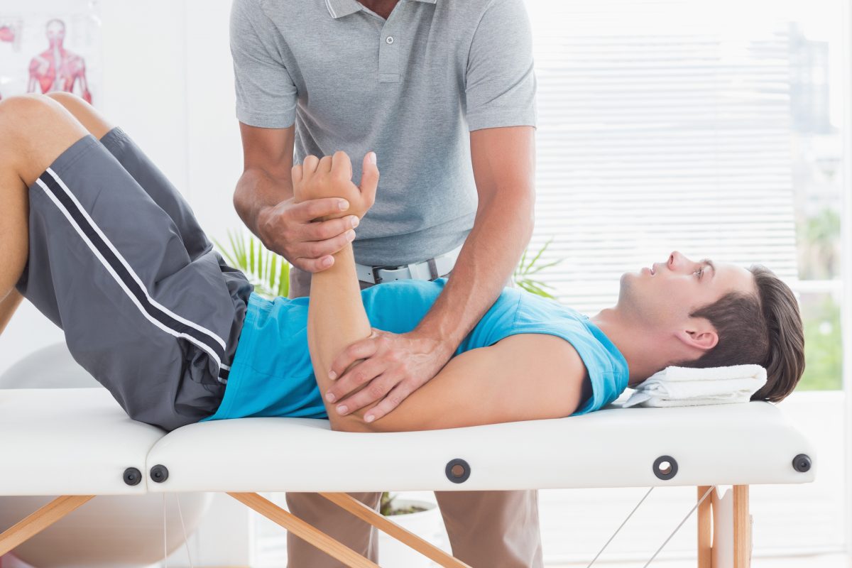
Diagnosing SCFE typically involves a combination of medical history review, physical examination, and imaging tests. The healthcare provider will inquire about the child's symptoms and perform a physical examination to assess the range of motion and stability of the hip joint.

Imaging tests are essential for confirming the diagnosis and determining the severity of the condition. X-rays are commonly used to visualize the position of the femoral head relative to the femoral neck. In SCFE, characteristic findings on x-rays include a slippage of the femoral head in relation to the neck, often described as a "ice cream slipping off a cone" appearance.

In some cases, additional imaging studies such as MRI or CT scans may be ordered to obtain more detailed images of the hip joint and surrounding structures.

Treatment Options for SCFE:

The primary goals of treatment for SCFE are to stabilize the hip joint, relieve symptoms, and prevent further slippage. The treatment approach may vary depending on the severity of the condition and the child's age and overall health. Common treatment options include:

  1. Surgical intervention: In most cases of SCFE, surgery is necessary to stabilize the hip joint and prevent further slippage. The two main surgical procedures used to treat SCFE are:

    a. In Situ Fixation: This involves inserting screws or pins into the femoral head and neck to hold them in place and prevent further slippage.

    b. Realignment Osteotomy: In severe cases or when the slip is chronic, a realignment osteotomy may be performed to reposition the femoral head and neck into the correct alignment.

  2. Non-surgical management: In some mild cases of SCFE, non-surgical measures such as rest, activity modification, and physical therapy may be recommended to alleviate symptoms and prevent worsening of the condition. However, close monitoring is essential to detect any progression of the slip.

Post-operative Care and Rehabilitation:

Following surgery, a period of immobilization and restricted weight-bearing may be necessary to allow the hip joint to heal properly. Physical therapy is an integral part of rehabilitation to improve strength, flexibility, and mobility of the hip joint. The rehabilitation process is tailored to each individual's needs and may continue for several months to achieve optimal recovery.

Long-term Outlook:

With prompt diagnosis and appropriate treatment, most children with SCFE can expect favorable outcomes and return to normal activities. However, untreated or poorly managed SCFE can lead to serious complications such as hip joint deformity, osteoarthritis, and chronic pain later in life. Regular follow-up appointments and monitoring are essential to detect any signs of recurrence or complications early on.

Conclusion:

Slipped Capital Femoral Epiphysis (SCFE) is a potentially debilitating hip disorder that primarily affects adolescents during their growth spurts. Early recognition of symptoms and timely intervention are crucial for preventing long-term complications and ensuring optimal outcomes. By understanding the causes, symptoms, and treatment options for SCFE, healthcare providers, parents, and caregivers can work together to provide the best possible care for children affected by this condition.

Tuesday, 5 March 2024

Orthopedic Screening of Newborns: Early Detection for Lifelong Health


Orthopedic screening of newborns plays a crucial role in identifying congenital musculoskeletal abnormalities early on, allowing for timely intervention and treatment. These screenings are vital for ensuring the proper development and function of a child's bones, muscles, and joints throughout their life.

During the initial examination shortly after birth, healthcare providers conduct a thorough assessment of the newborn's musculoskeletal system. This assessment includes a visual inspection and palpation of the baby's limbs, spine, and joints to detect any abnormalities or asymmetries. Additionally, specific orthopedic maneuvers may be performed to assess for conditions such as hip dysplasia or clubfoot.

One of the most common conditions screened for in newborns is developmental dysplasia of the hip (DDH). DDH occurs when the hip joint fails to develop properly, potentially leading to long-term issues with mobility and function if left untreated. Early detection through orthopedic screening allows for non-invasive interventions such as harnesses or braces to properly align the hip joint and promote normal development.

Another condition that may be identified during newborn orthopedic screening is clubfoot, a congenital deformity characterized by inwardly twisted or rotated feet. Prompt intervention with techniques such as the Ponseti method, involving gentle manipulation and casting of the foot, can effectively correct clubfoot and prevent future complications.

In addition to these specific conditions, orthopedic screening also helps identify general musculoskeletal abnormalities such as limb length discrepancies, joint contractures, or spinal deformities. Early detection of these issues allows for appropriate referrals to pediatric orthopedic specialists for further evaluation and management.

The benefits of orthopedic screening extend beyond infancy, as timely intervention can prevent long-term disability and improve overall quality of life for affected children. By addressing musculoskeletal abnormalities early on, healthcare providers can minimize the need for extensive treatments or surgeries later in life.

Furthermore, orthopedic screening serves as an essential component of comprehensive newborn care, ensuring that all aspects of a child's health are thoroughly evaluated from the very beginning. Early identification of musculoskeletal issues enables healthcare providers to collaborate with families to develop personalized treatment plans and provide ongoing support throughout the child's development.

It's important to note that while orthopedic screening is a routine part of newborn care in many healthcare settings, not all infants will require intervention. However, for those who do, early detection and appropriate management can make a significant difference in their long-term outcomes.

In conclusion, orthopedic screening of newborns plays a critical role in identifying and addressing musculoskeletal abnormalities early in life. By detecting conditions such as DDH, clubfoot, and spinal deformities at birth, healthcare providers can initiate timely interventions that promote optimal development and function. Through comprehensive newborn care that includes orthopedic screening, we can ensure the lifelong health and well-being of every child.

Friday, 29 December 2023

Understanding Various Aspects of Bow Legs Treatment

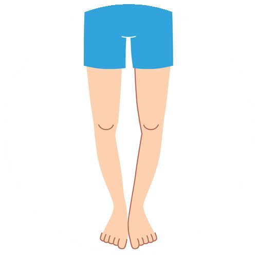
Each infant is born bowlegged as he remained in mother’s womb in a folded position. Hence, bow legs are considered normal in children whose age is less than 18 months. As the child starts walking, his legs get straightened gradually. But bow legs are considered as a physical deformity if it is present when the child is about 3 years old. As a form of physical deformity, bow legs or genu varum is marked by bowing of the child’s lower leg in relation to his thigh.

Bow Legs | Children's Podiatrist | Melbourne Podiatrists & Orthotics

Bow legs may be caused due to a variety of illnesses. Normally, the physical deformity is caused due to abnormal development of bones or fractures that have not healed properly. However, bow legs are also caused due to Blount’s disease, or lead or fluoride poisoning. Sometimes the physical deformity is caused due to rickets, an illness caused due to lack of Vitamin D.

The parents can identify the physical deformity based on a number of symptoms. They can know if the child is bowlegged when his knees to not touch while standing with both feet together. Also, a bowlegged child can bow his symmetrically on both sides of his body. The parents must avail the assistance of a seasoned orthopaedic immediately if bow legs continue in a child whose age is more than 3 years.

A seasoned child orthopaedic can diagnose bow legs simply by observing the child’s knees. Some healthcare providers even diagnose bow legs by measuring the distance between two knees of the child when he lies on the back. However, the orthopaedic also perform blood test to check if the deformity is caused due to rickets. They even recommend x-rays when the bowing is extreme or based on the test results. But x-ray is recommended only if the child is older than 3 years.

The seasoned orthopaedics monitors the growth of the bow legged child constantly, and observe his knees once in 6 months. Also, they recommend treatment for bowleg only when the child’s condition is extreme. Many orthopaedics treat bow legs through special braces, casts, or shoes. The skilled orthopaedics also corrects the deformity in an adolescent child by performing bow legs surgery. However, the bow legs treatment varies based on the specific condition of each child.

The parents do not have any option to prevent bow legs. Hence, they must avail the assistance of a skilled orthopaedic to monitor the child’s condition and prevent his condition from becoming extreme. The orthopaedic will further recommend the best way to treat bow legs on time and avoid chances of developing arthritis in the knees or hips. Also, the parents must choose a skilled and experienced orthopedic to avail modern treatment, and correct the physical deformity with minimal scar and morbidity.

Thursday, 30 November 2023

What is the most common surgery for an orthopedic surgeon?

Pediatric orthopedics is a specialized branch of orthopedic medicine that focuses on the diagnosis and treatment of musculoskeletal conditions in children, from infancy through adolescence. Pediatric orthopedic surgeons are medical doctors who have completed additional training in orthopedic surgery with a specific focus on children.

Children’s bones, muscles, and joints are still growing and developing, which can lead to unique orthopedic conditions and challenges. Pediatric orthopedic surgeons are experts in managing these conditions and providing appropriate treatment options for children.

Blog | Kids Orthopedic Clinic

Some common conditions that pediatric orthopedic surgeons treat include:

  1. Congenital anomalies: These are conditions that children are born with, such as clubfoot, hip dysplasia, limb length discrepancies, and scoliosis.
  2. Fractures and trauma: Children are prone to fractures due to their active lifestyles. Pediatric orthopedic surgeons are skilled in treating fractures and ensuring proper healing.
  3. Sports injuries: Children and adolescents who participate in sports are susceptible to sports-related injuries, including sprains, strains, fractures, and ligament tears. Pediatric orthopedic surgeons can diagnose and treat these injuries, often with a focus on preserving future joint function.
  4. Musculoskeletal infections: Children may develop infections in their bones or joints, such as osteomyelitis or septic arthritis. Pediatric orthopedic surgeons work closely with infectious disease specialists to provide appropriate treatment.
  5. Neuromuscular conditions: Certain neurological conditions, such as cerebral palsy or spina bifida, can affect a child’s musculoskeletal system. Pediatric orthopedic surgeons collaborate with other specialists to develop comprehensive treatment plans for these patients.
  6. Growth-related conditions: Children may experience conditions related to abnormal growth, such as limb length discrepancies or angular deformities. Pediatric orthopedic surgeons can assess and manage these conditions through various treatment options, including surgery, bracing, or physical therapy.

shutterstock_262417277-e1523306430412

Pediatric orthopedic surgeons often work in collaboration with other healthcare professionals, including pediatricians, physical therapists, occupational therapists, and rehabilitation specialists, to provide comprehensive care for children with orthopedic conditions.

Tuesday, 31 October 2023

What are overuse injuries in the pediatric population?

Overuse injuries in pediatrics refer to a category of injuries that occur when a child or adolescent repeatedly engages in a specific physical activity or sport, placing excessive stress on a particular part of their body. These injuries are common among young athletes who train intensively or participate in a single sport year-round. Overuse injuries can lead to pain, impaired performance, and potentially long-term musculoskeletal issues. Here are some key points to understand about overuse injuries in pediatrics:

  1. Types of Overuse Injuries:Overuse injuries can affect various parts of the body, including:
  • Growth Plate Injuries: The growth plates, areas of developing tissue near the ends of bones, are vulnerable to overuse injuries.
  • Stress Fractures: These are small cracks or fractures in bones due to repetitive impact.
  • Tendonitis: Inflammation of tendons caused by repetitive movements.
  • Muscle Strains: Overuse can lead to strains or tears in muscles.
  • Apophysitis: Inflammation of the bony attachment points of muscles, commonly seen in areas like the heel (Sever’s disease) or knee (Osgood-Schlatter disease).
  1. Risk Factors:Several factors increase the risk of overuse injuries in pediatric athletes, including:
  • Specialization: Young athletes who specialize in a single sport and train intensively year-round are more susceptible to overuse injuries.
  • Growth and Development: Rapid growth during puberty can change the biomechanics of a child’s body, making them more prone to injury.
  • Inadequate Rest: Lack of adequate rest and recovery between activities or seasons can contribute to overuse injuries.
  1. Symptoms:The symptoms of overuse injuries may include pain, swelling, tenderness, reduced range of motion, and a decline in athletic performance. Children may also experience pain that worsens with activity and improves with rest.
  2. Prevention and Management:Preventing overuse injuries in pediatrics involves a combination of strategies:
  • Monitoring Load: Coaches, parents, and athletes should be vigilant about monitoring training loads to ensure they are appropriate for the child’s age and development.
  • Periodization: Training should incorporate periods of rest, cross-training, and reduced intensity to allow the body to recover.
  • Proper Technique: Teaching young athletes correct techniques and movement patterns can help reduce the risk of overuse injuries.
  • Adequate Rest: Children should have scheduled rest days and avoid year-round intensive training in a single sport.
  • Early Intervention: If an overuse injury occurs, early intervention, including rest, physical therapy, and sometimes bracing, is essential to prevent long-term consequences.
  1. Importance of Multisport Participation:Encouraging children to participate in multiple sports or activities can reduce the risk of overuse injuries by promoting a more balanced use of their bodies and reducing the strain on specific muscle groups and joints.

In conclusion, overuse injuries in pediatrics are a significant concern, especially among young athletes. Preventing these injuries involves a combination of strategies, with a focus on proper training, rest, and early intervention to protect the growing bodies of children and adolescents.


Tuesday, 3 October 2023

Identifying and Treating Neglected Fracture in Kids


According to the Canadian Paediatric Society, “Fractures are common injuries in childhood. While most fractures are caused by accidental trauma, inflicted trauma (maltreatment) is a serious and potentially unrecognized cause of fractures, particularly in infants and young children.

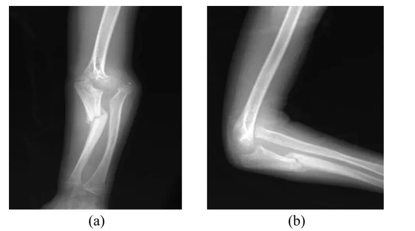
Fractures in children occur when excessive force is applied to the bone. As the bone cannot absorb excessive force, broken bones or fractures occur. Excessive force can cause multiple fractures or specific fractures like wrist fracture, elbow fracture, or femur fracture.

Also, fractures in kids occur due to a variety of reasons – trauma, falls, direct hit, injuries, and lack of calcium. You cannot control or change the causes and number of fractures in your child. But you must treat the neglected factors early and properly to keep your child fit by eliminating the chances of functional disability and permanent deformity.

Screenshot_9

What Should You Know to Treat a Neglected Fracture in Your Child Successfully?

Symptoms

A neglected fracture might occur in your child due to a variety of reasons. Also, the symptoms of neglected broken bones vary from one child to another. You can identify neglected fractures based on common systems like pain, swelling, bruising, redness, and warmth. Also, the affected area sometimes appears deformed due to the broken bone. It is always important to consult a pediatric orthopedic surgeon once your child finds it difficult to use or move the injured bone normally and naturally.

Diagnosis

Experienced pediatric orthopedic surgeons diagnose neglected fractures in a variety of ways. They always start diagnosing by inspecting the affected area closely and physically. The physical inspection, along with the child’s medical history, helps experienced orthopedic surgeons to identify the condition and causes of the fracture. However, it is also common for orthopedic surgeons to identify the cause of broken bones by recommending X-rays, CT scans, MRI scans, and similar imaging examinations.

Treatment Options

The orthopedic surgeons diagnose neglected fractures in a variety of ways according to the severity and symptoms of the broken bone. Also, they prepare a personalized treatment plan for the neglected fracture based on your child’s age, medical history, and severity of the fracture. Also, they keep in mind your child’s capacity to tolerate specific medication or treatment options.

Screenshot_10

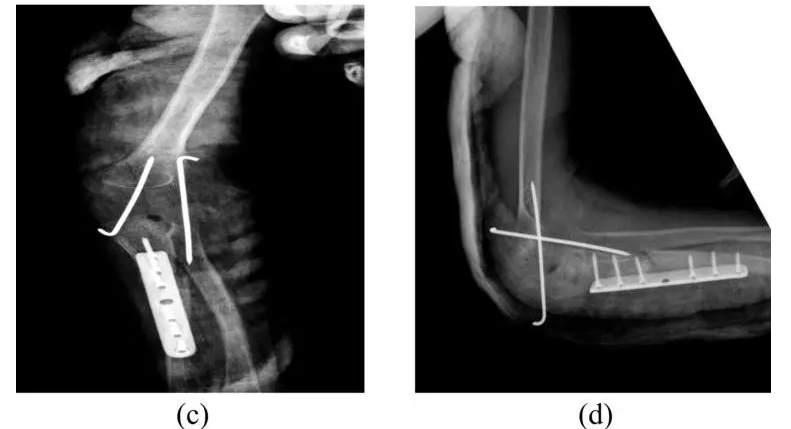
Experienced pediatric orthopedic surgeons always start treating neglected fractures through non-surgical procedures like medication, cast/splint, and traction. But they treat neglected fractures surgically when the non-medical procedures do not work. They perform surgeries to reposition the broken bones or implant fixation devices.

Recovery

The non-medical procedures treat neglected fractures gradually over a period of time. Your child will become fit and active after taking medication or wearing the cast/splint regularly for a specific duration. However, you must get the affected area inspected by the orthopedic surgeon regularly according to the treatment plan. On the other hand, your child needs to rest the affected bone completely and follow operation-care recommended by the surgeon when the neglected fracture is treated surgically.

You can easily get a neglected fracture in your child diagnosed accurately and treated effectively by consulting an experienced pediatric orthopedic surgeon. The orthopedic surgeons will help you to eliminate the risk of functional disability and permanent deformity by treating neglected fractures accurately and successfully.

Thursday, 14 September 2023

Recognizing Signs of Cerebral Palsy in a Child

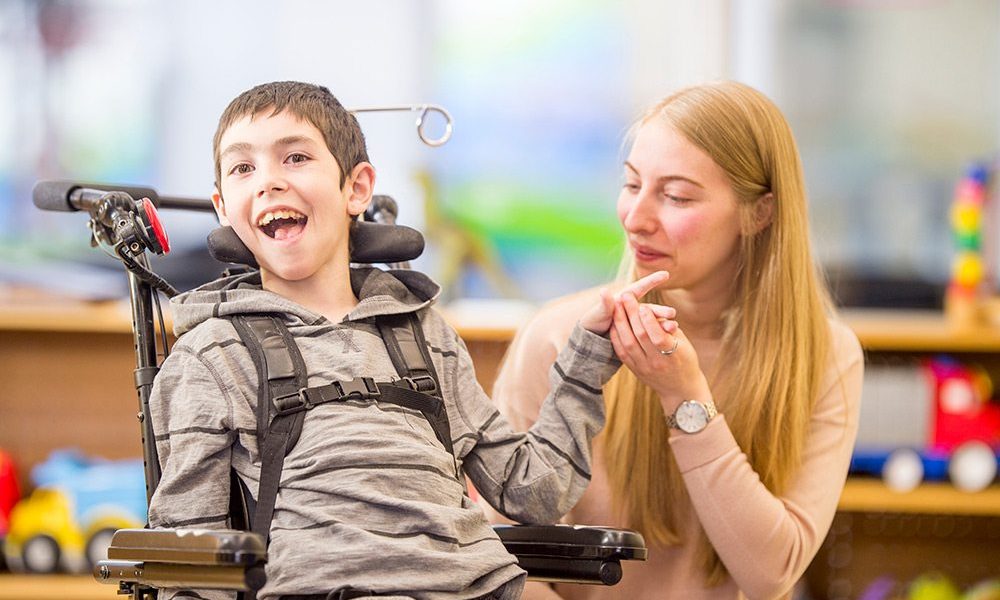
As a permanent and complex disorder, cerebral palsy is caused due to damage caused to the brain of a child. The brain damage can occur due to a number of reasons. Also, the damage may be caused to the child’s brain before, during or after his birth. At present, there is no permanent cure for cerebral palsy. The seasoned orthopedics tries to reduce the symptoms and effects of the permanent disorder through medication, orthotic devices, speech therapy, physical therapy or occupational therapy.

Cerebral-Palsy-1000x600

Simple Ways to Care for a Child with Cerebral Palsy

It is always important for parents to get their kids diagnosed and treated for cerebral palsy immediately. Also, they must understand various aspects of cerebral palsy management to make the child feel comfortable. But cerebral palsy is often diagnosed only after 18 months of the child’s birth.  There are many instances when parents failed to diagnose symptoms of mild cerebral palsy when the kid is 3 to 5 years old. That is why; it become essential for parents to know how to recognize the signs of cerebral palsy in a kid.

To identify the developmental disabilities, the parent must understand know four distinct categories of developmental progress of a child. Broadly, the developmental progress of a child of a child can be divided into four categories – physical, social, cognitive and emotional. Also, the parent must know the normal stages of development to identify the developmental issues immediately. However, the parents must get the child diagnosed for cerebral palsy when they notice lack of physical development.

The parents must get the child diagnosed for cerebral palsy when the child does not meet common developmental milestones like crawling, sitting, walking, rolling over, visual alertness, and picking of small objects. The orthopedic will observe the development of the child over a specific period of time and screen the child regularly to identify improvement disabilities. He will further observe and assess the development of the child based on his posture, muscle tones and reflexes. However, the severity and symptoms of cerebral palsy differ from one child to another.

The child with cerebral palsy further experiences symptoms of the disorder according to the location of movement problems. The symptoms further get aggravated if the child is not provided with adequate care and effective treatment on time. Hence, it is important for parents to get the child diagnosed for cerebral palsy immediately and promptly. At the same time, they also need to ensure that the child is diagnosed and treated by a skilled and experienced orthopedic to manage and reduce symptoms of the complex disorder over a longer period of time.

Know more about stay social with us on: Facebook .